Fig. S8.
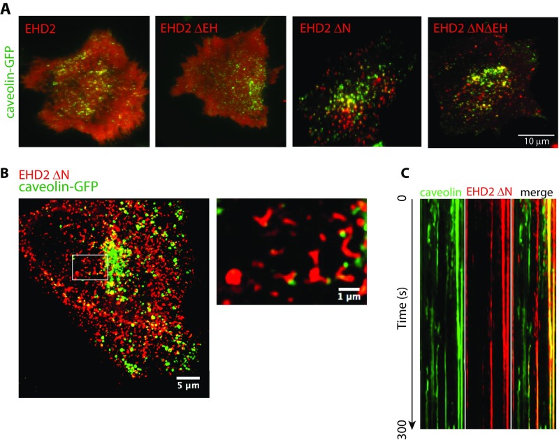
Deletion of the N terminus of EHD2 affects caveolae stability and morphology, related to Fig. 5. (A) Florescence micrographs that represent the first frame in a live-cell TIRF experiment where caveolin 1-GFP (green) and indicated EHD2 variants (red) are coexpressed. (B) Representative maximum projection image of a caveolin 1-GFP FlpIn cell that transiently expresses EHD2 ΔN. The image was captured by Airyscan confocal microscopy, and the Inset in the image to the Right shows the magnification of the indicated area. (C) Kymograph from live-cell TIRF imaging of a cell showing the movement of colocalized (yellow) and individual caveolin 1 or EHD2 ΔN assemblies at the cell surface during a 300-s movie.

