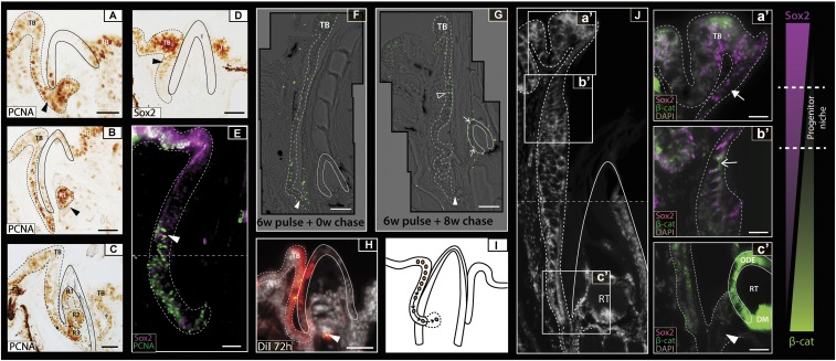
Fig. 2.
Localization of a dental progenitor cell niche within the pufferfish dental lamina. (A–C) P. baileyi PCNA immunohistochemistry reveals high levels of cellular proliferation within the oral epithelium. As replacement teeth progress from late-initiation (A) to morphogenesis (C), the new tooth generation (R1) buds from the dental lamina (B). Successive rounds of replacement show that the dental generations stack on one another within an enameloid outer casing (black line) (C). (D) Sox2 immunohistochemical labeling during dental replacement initiation depicts high levels of Sox2 within both the developing taste buds (TB) and the dental progenitor site located within the labial oral epithelium (dental lamina) (black arrowhead). (E) Double immunofluorescence treatment for Sox2/PCNA in T. niphobles shows low levels of PCNA expression within the Sox2+ cells of the presumptive dental progenitor niche and cells within the aboral dental lamina exhibiting high levels of PCNA. The horizontal dashed line depicts image stitching of two adjacent images. White arrowhead marks region of overlapping PCNA/Sox2 expression. (F) BrdU pulse/chase experiments (0.2 mM) show the incorporation of BrdU into dividing cells after 6 wk of treatment, with high levels of incorporation noted in the distal dental lamina next to the base of the beak (white arrowhead). (G) After a further 8-wk chase, label-retaining cells were found in the most superficial dental lamina cells (open arrowhead) but not in the distal dental lamina (white arrowhead). Label-retaining cells found in the dental epithelium of the developing tooth are indicated by a white arrow. Images in F and G are composites of multiple images taken at high magnification and stitched together. (H) DiI labeling of the labial oral epithelium in P. suvattii highlighted this region as a presumptive source of dental progenitor cells. DiI was detected within the outer dental epithelium of the tooth (white arrowhead) 72 h post DiI treatment. (I) As summarized in a schematic representation, we observed a continuous field of Sox2+ cells between the labial taste bud and the dental progenitor site, with cells from the latter migrating and contributing to the new dental generations. Black arrows represent the direction of cell movement. (J) Sox2/ABC double immunohistochemical labeling on adult C. travancoricus highlights epithelial Sox2+/ABC− (a′, white filled arrow), Sox2+/ABC+ (b′, white arrow), and Sox2−/ABC+ (c′, white arrowhead) regions within the dental lamina. Coexpression of these markers marks the site of activation of putative dental progenitors within the oral epithelium. Dashed line across (J) depicts image stitching of two adjacent images. Images are orientated with labial to the left and oral to the top. The dotted line in all images depicts the boundary of the oral epithelium and the end of the dental lamina. DM, dental mesenchyme; ODE, outer dental epithelium; R1–3, replacement tooth generations; RT, regenerating tooth; TB, labial taste bud. (Scale bars: 25 µm in A–E; 20 µm in F, a′–F, c′; 50 µm in G and H; 15 µm in I.)

