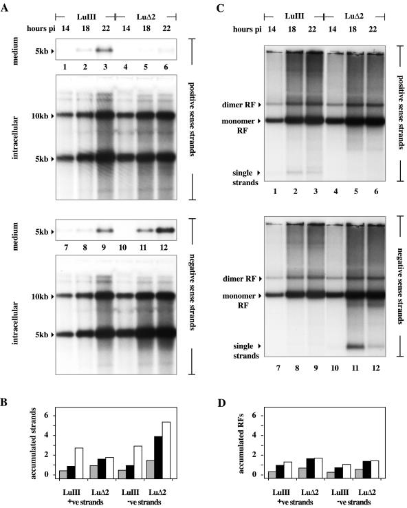
FIG. 5.
Accumulation of unit length viral DNA in cells infected with the LuIII and LuΔ2 viruses. (A) Southern transfers of total viral DNA either released into the medium or retained in cells 14, 18, and 22 h postinfection (pi) following electrophoresis through denaturing gels, transfer, and incubation with strand-specific probes. Total unit length DNA was quantitated by PI analysis against standard DNAs run in the same gel (as detailed in the text). (B) Accumulation of unit length (5- plus 10-kb) DNA strands of each polarity with time, expressed on the y axis as micrograms of DNA quantitated for each strand following electrophoresis through denaturing gels. Individual histogram blocks shaded gray or black or unshaded represent DNA harvested at 14, 18, and 22 h postinfection, respectively. (C) Southern transfers of total viral DNA from cells 14, 18, and 22 h postinfection, following electrophoresis through neutral agarose gels. The migration positions of monomer RF, dimer RF, and progeny single-stranded DNAs are indicated. (D) Accumulation of unit length duplex DNA (5 plus 10 kb) with time, expressed on the y axis as micrograms of DNA quantitated for each strand following electrophoresis through neutral gels. Individual histogram blocks shaded gray or black or unshaded represent DNA harvested at 14, 18, or 22 h postinfection, respectively. +ve, positive; −ve, negative.

