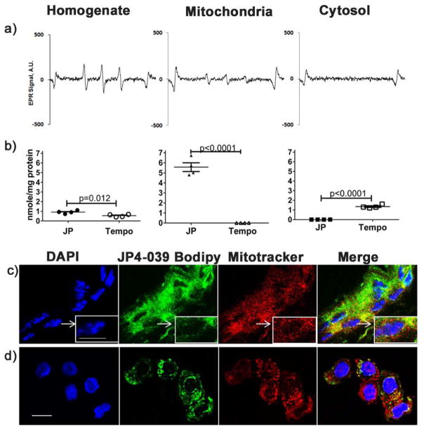
Figure 1. Topical JP4-039 localizes to keratinocyte mitochondria.
Whole skin homogenates and subcellular fractions were derived from mouse skin 30 min after topical application of JP4-039 or Tempo. Typical EPR spectra of JP4-039 nitroxide radical in unfractionated cell homogenates and mitochondrial and cytosolic subfractions are shown (a). EPR spectra were used to quantitate levels of nitroxide radicals present in each fraction in JP4-039 or Tempol treated skin (n=4) (b). JP4-039 bodipy treated intact skin (c) and isolated keratinocytes (d) were labeled with Mitotracker Red-CMXROS and Dapi and imaged by confocal microscopy. Merged images confirm localization of JP4-039-Bodipy in the mitochondria (bar=10μm. Statistical significance was determined using a t-test.

