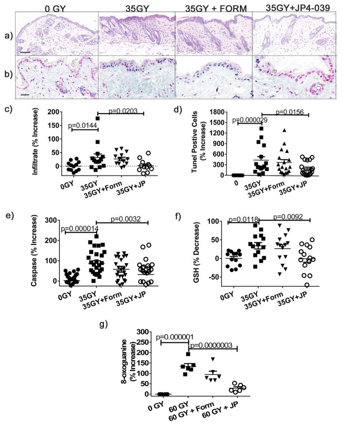
Figure 3. Topical JP4-039 prevents radiation induced cutaneous apoptosis, inflammation, antioxidant depletion, and DNA damage.
35 Gy irradiated mice were treated or not 0.5h after irradiation. Shown are representative H&E sections 4 h after irradiation (bar = 40μm) (a), quantified cellular infiltrates (b), TUNEL stained sections (b) with positive cells staining blue and negative cells red (bar = 20μm), quantification of TUNEL positive cells (d), and Caspase 3/8 levels determined in homogenates (e). GSH levels were determined enzymatically from skin homogenates (f). DNA damage was evaluated by 8-oxoguanine staining and expressed as percent positive cells (g). Results are presented as % increase (((treated/untreated average)−1)*100). Data are presented as mean ± SEM (n=13–22). Statistical significance was determined by ANOVA followed by a Bonferroni post-test with significance set at p<0.05.

