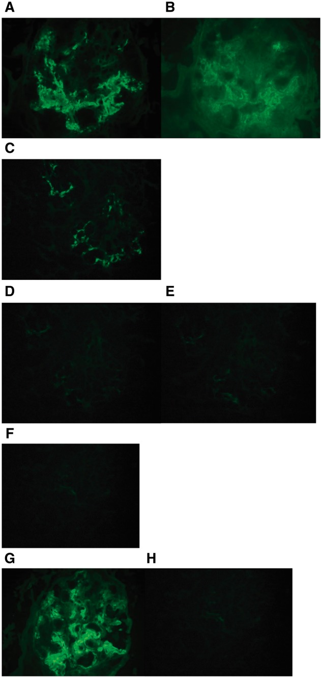
Fig. 2.

Immunofluorescence microscopy showed granular mesangial and occasional capillary wall staining for (A) IgG and (B) C1q. IgG subclasses showed positive glomerular staining for (C) IgG1 only, whereas the glomeruli were negative for (D) IgG2, (E) IgG3 and (F) IgG4. Additionally, the glomeruli showed positive staining for (G) kappa light chain, but were negative for (H) lambda light chain (original magnification, 400×).
