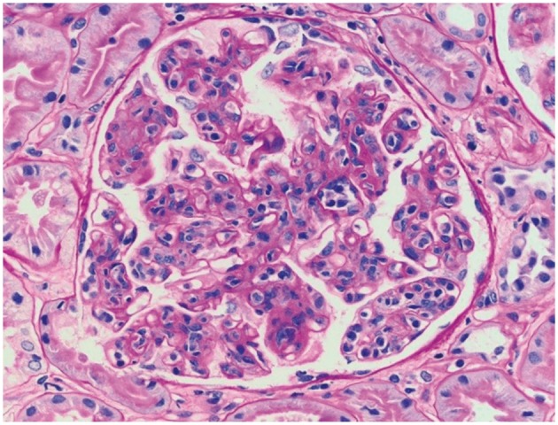
Fig. 4.

Proliferative GN with monoclonal IgG deposits in the kidney allograft biopsy specimen from patient 2, 9 months posttransplantation. Light microscopy showed features of membranoproliferative GN, including accentuated lobulation of the capillary tufts, mesangial and endocapillary hypercellularity, and thickening and duplication of the glomerular basement membranes (periodic acid–Schiff stain; original magnification, 400×).
