
Figure 3.
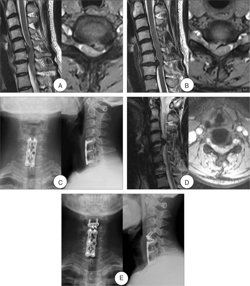
A 40-year-old woman developed one-level ASD 3 years after initial surgery. (A) and (B) Preoperative MRI of this patient shown severe compressions of the spinal cord at C5-6 and C6-7 levels. (C) Radiograph after initial surgery shown ACDF at C5-6 and C6-7. (D) MRI at 3-year follow-up shown development of ASD and spinal cord compression at C4–5. (E) Radiograph after the reoperation shown ACDF with the Zero-profile device at C4-5 and the fixed plate of the initial surgery was not removed. ACDF = anterior cervical decompression and fusion, ASD = adjacent segment disease, MRI = magnetic resonance imaging.