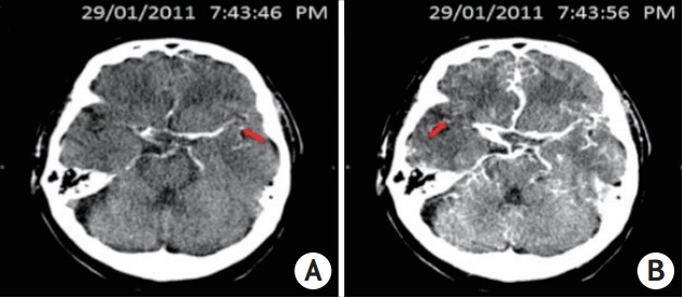
Figure 1.

This picture showed a typical case of relative filling time delay (rFTD): a 71-year-old male patient presenting with left hemiplegia and dysphasia arrived hospital and underwent computer tomography examination 81 minutes after onset. Computer tomography angiography indicated the occluded right internal carotid artery and M1 segment of the right middle cerebral artery. Computer tomography perfusion source imaging (CTP-SI) illustrated that contrast signal (red arrow) firstly appeared in the normal side’s Sylvian fissure in 7:43:46 PM (A), whereas showed up in the occlusion side’s Sylvain fissure in 7:43:56 PM (B), therefore, the time gap of 10 s between bilateral contrast appearances in Sylvian fissure was identified as an rFTD.
