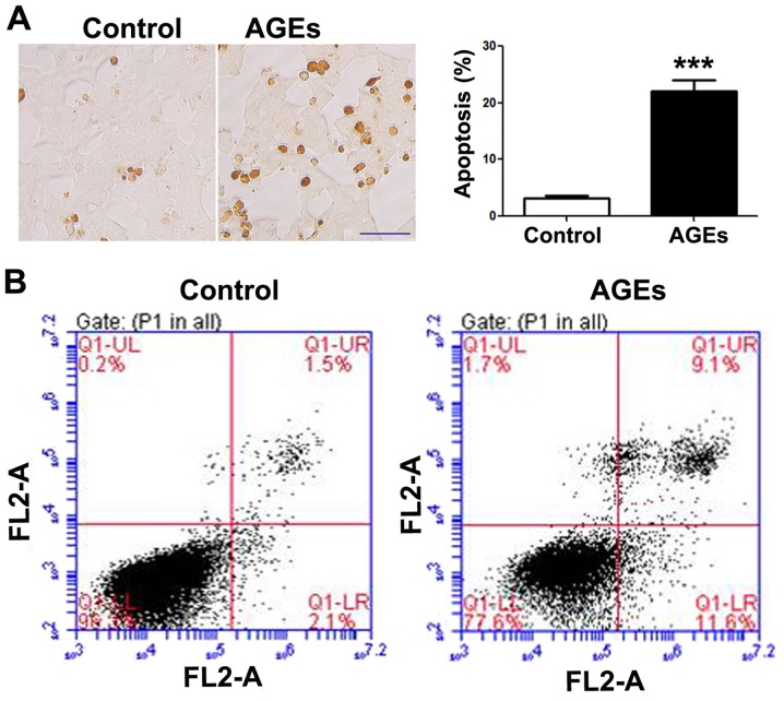
Figure 1.
Advanced glycation end products (AGE) induce the apoptosis of INS-1 cells. (A) TUNEL staining detected the apoptosis of INS-1 cells 48 h following exposure to AGEs (200 µg/ml). Data are presented as the means ± SEM (n=3). ***P<0.001, compared with the control group. Scale bar, 50 µm. (B) Flow cytometry was used to detect the apoptosis of INS-1 cells. INS-1 cells were exposed to 200 µg/ml AGEs for 48 h, followed by Annexin V/PI staining assay to detect the apoptosis of INS-1 cells by flow cytometry. Top left quadrant, necrotic cells; bottom left quadrant, live cells; top right quadrant, late apoptotic cells; bottom right quadrant, early apoptotic cells.

