Figure 1.
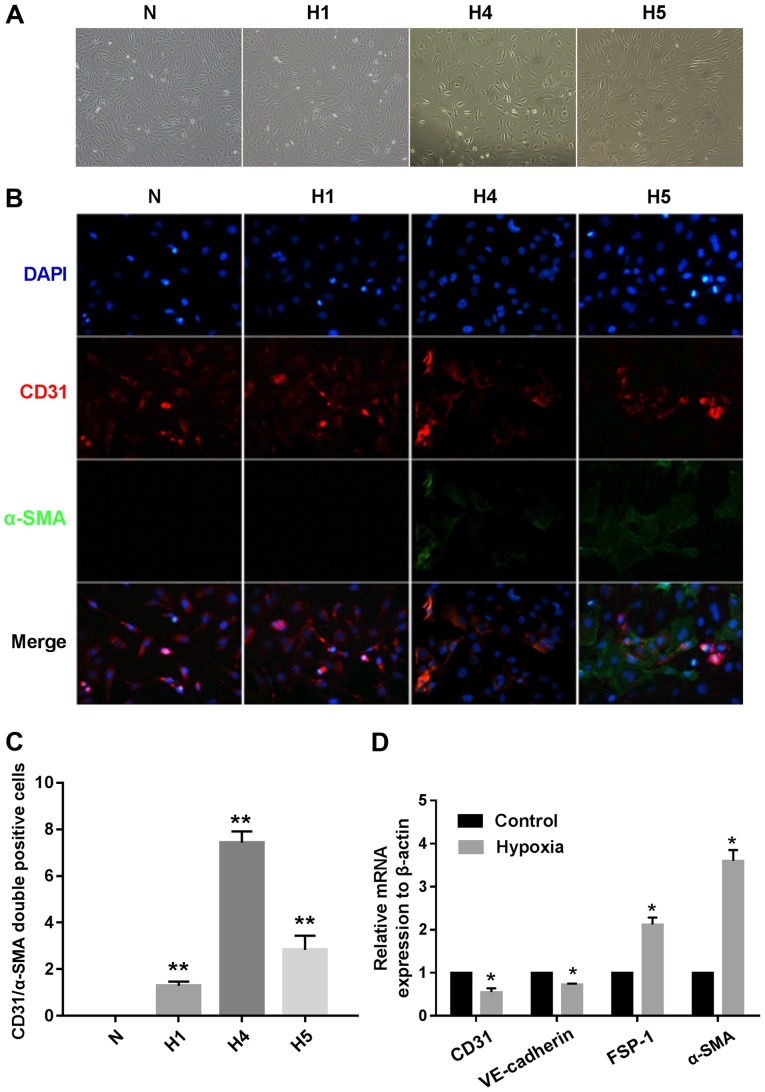
Hypoxia induces human cardiac microvascular endothelial cells (HCMECs) to mesenchymal transition. HCMECs were cultured under either normoxic conditions (N) or hypoxic conditions for 1, 4 and 5 days, respectively (H1, H4 and H5). (A) Representative phase contrast light microscopy images showing morphological changes in HCMECs (x100 original magnification). (B) Double immunofluorescence staining with antibodies to CD31 (red) and α-smooth muscle actin (α-SMA) (green). Nuclei were counterstained with DAPI (blue). Acquisition of a spindle-shaped morphology upon hypoxia was correlated with α-SMA acquisition and loss of CD31 (x200 original magnification). (C) Graph indicates quantification of CD31+/α-SMA+ cells from each group. Bars indicate mean ± SD, n=7 independent experiments. **P<0.01 vs. normoxia group. (D) RT-PCR data showing the mRNA expression levels of the endothelial markers (CD31 and VE-cadherin) and mesenchymal markers [fibroblast-specific protein (FSP)-1 and α-SMA] in the normoxia and hypoxia groups. Results were normalized to that of reference gene β-actin (n=7 independent experiments; *P<0.05 vs. normoxia group).

