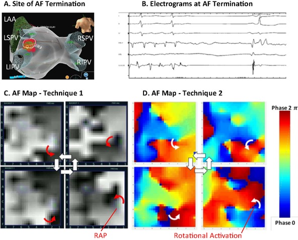
Figure 3.

Identification of rotations at site of termination by both techniques (patient ID 2) in a 72‐year‐old man with persistent AF. (A) Prospective guided ablation at the carina of left pulmonary vein (B) terminated persistent AF prior to PVI. (C) AF snapshots from technique 1 show counterclockwise rotation at termination site CD2 for >10 cycles particularly in the second half of movie 3. (D) AF snapshots from technique 2 also show counter clockwise activation at this termination site (movie 4). Complex fibrillatory activity and competing wavefronts are also seen.
