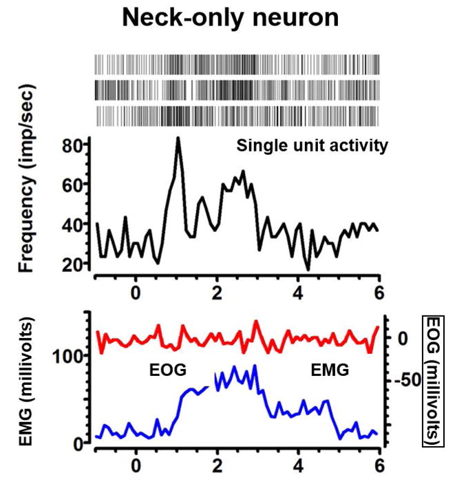
Fig 3.

An example of neck only neuron depicting increase in the firing rate (black trace) with isometric neck muscle (sternocleidomastoid) contraction (blue traces) without coactivation of eye movements (red trace). Inset of the panel depicts raster plot, each marker of the raster depicts the presence of spike at a given time.
