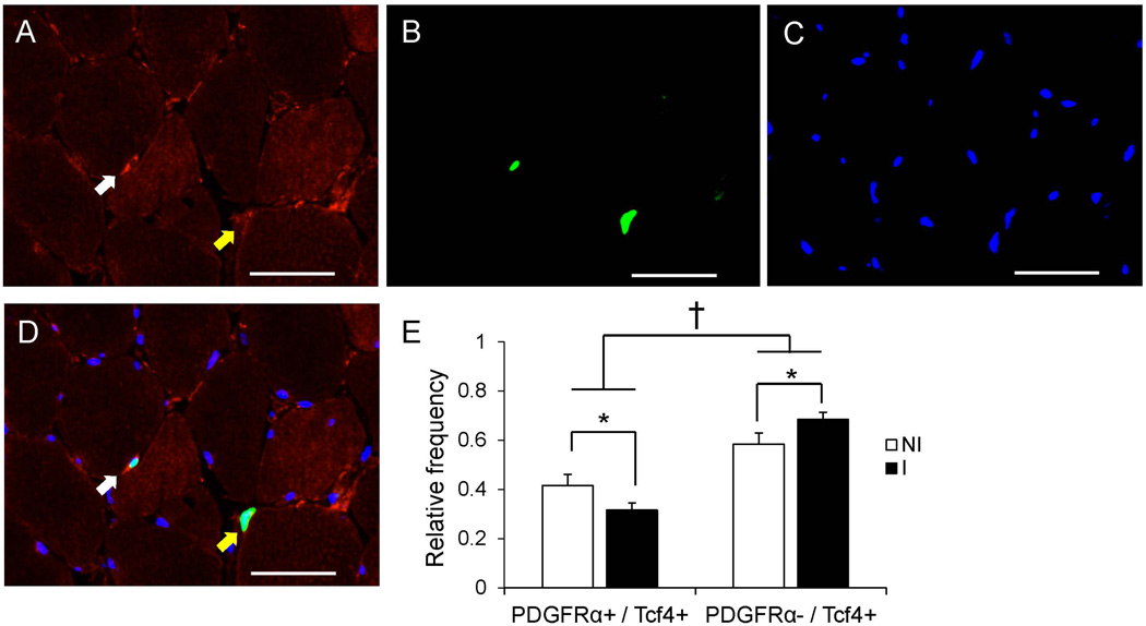
Figure 4. PDGFRα+ and Tcf4+ co-localization is affected by ACL injury.
Representative images of PDGFRα (A), Tcf4 (B) and DAPI (C) staining in ACL injured muscle. (D) Merged immunohistochemical image demonstrating a PDGFRα+ / Tcf4+ cell (white arrow) and a PDGFRα− / Tcf4+ cell (yellow arrow). Scale bar = 50µm. (E) Quantification presented as mean relative frequency of cells staining for both PDGFRα and Tcf4 versus Tcf4 alone ± SEM. * Significantly different from the non-injured limb (p < 0.05); † Significant effect of PDGFRα-staining status (p < 0.05).

