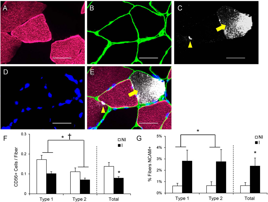
Figure 7. Reduced satellite cell abundance and increased frequency of muscle fiber denervation following ACL injury.
(A–D) Representative images of myosin heavy chain type 1 (A), laminin (B), CD56/Neural Cell Adhesion Molecule (NCAM) (C) and DAPI (D) staining in ACL injured muscle. (E) Merged immunohistochemical image demonstrating a CD56+ satellite cell (yellow arrowhead) and a NCAM+ muscle fiber (yellow arrow). Scale bar = 50µm. (F) Quantification of fiber type-specific and pooled CD56+ satellite cell content, expressed as mean CD56+ satellite cells per fiber ± SEM. (G) Quantification of fiber type-specific and pooled NCAM+ fibers, expressed as mean percentage of fibers positive for NCAM ± SEM. N = 10 subjects. NI = non-injured; I = injured. * Significantly different from the non-injured limb (p < 0.05); † Significant effect of fiber type (p < 0.05).

