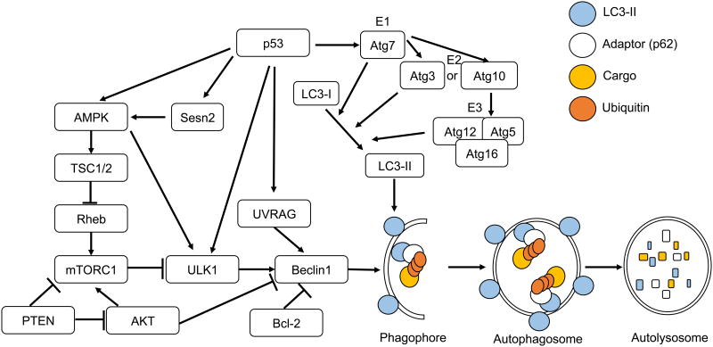
Figure 1. Mechanisms of Autophagy.
Autophagy is initiated upon activation of the AMPK signaling pathway. AMPK can be activated by p53 and Sesn2 in response to cellular stress. AMPK then activates TSC1/2 to inhibit mTORC1. mTORC1 is further negatively regulated by PTEN to activate autophagy. ULK1 is activated by inhibition of mTORC1, direct phosphorylation by AMPK, or p53-mediated transcriptional upregulation. ULK1 then forms a complex which activates Beclin1. Beclin1, which binds to Bcl-2 in normal conditions, dissociates from Bcl-2 and binds to UVRAG. UVRAG, a p53 target gene, promotes the formation of the Beclin1 complex to initiate phagophore nucleation. Two ubiquitin-like conjugation systems facilitate the lipidation of LC3-I to form LC3-II, and elongation of the burgeoning phagophore. E1-like activating enzyme Atg7 leads to the activation of E2-like enzyme Atg3 (System 1) or Atg10 (System 2), subsequently the activation of the E3-link enzyme complex Atg5-Atg12 conjugate with Atg16, and subsequently the generation of LC3-II. LC3-II binds to an adaptor protein or autophagy receptor, such as p62, which in turn binds to cargo with modifications such as polyubiquitination. The autophagosome encloses the cargo and subsequently fuses with a lysosome to form an autolysosome. Adaptor and cargo are degraded at the autolysosome and the resulting molecules are recycled to the cytoplasm.

