
Figure 4.
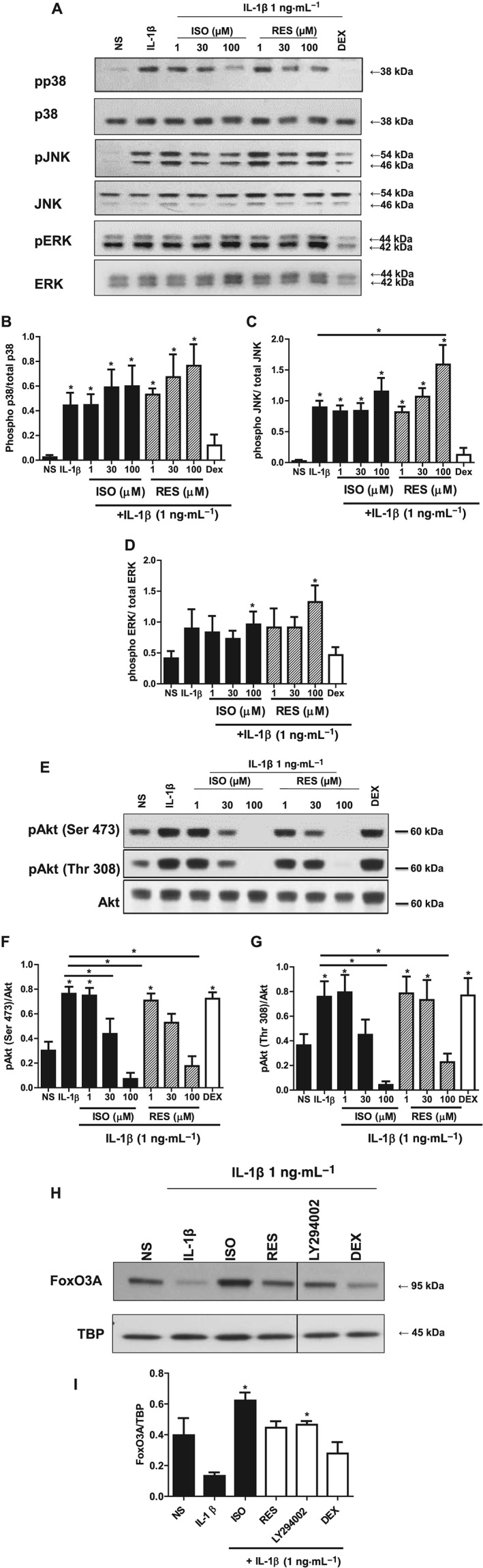
Effects of stilbene analogues and dexamethasone (DEX) on the major intracellular inflammatory signalling pathways. (A) Representative blots and quantification of phosphorylated and total p38 (B), JNK (C) and ERK (D) protein expression in A549 cells treated with various concentrations of either isorhapontigenin (ISO) or resveratrol (RES) (1–100 μM) or 1 μM DEX for 1 h before stimulation with 1 ng·mL−1 IL‐1β for 40 min. IL‐1β induced increase in levels of phosphorylated p38, JNK and ERK protein expression, which were suppressed by DEX but not stilbene analogues (n = 4). (E) Representative blots and quantification of phosphorylated Ser473 (F) or Thr308 (G) and total Akt protein expression in A549 cells treated with various concentrations of either ISO or RES (1–100 μM) or 1 μM DEX for 1 h before stimulation for 40 min. IL‐1β induced increase in levels of phosphorylated Akt (at Ser473 and Thr308) protein expression, which were suppressed by stilbene analogues and not by DEX (n = 5). (H) Representative blot of nuclear protein expression of FoxO3A and densitometry (I) showing reduction with IL‐1β at 60 min, and this reduction was prevented by ISO, RES and LY294002 but not DEX (n = 3). Data are mean ± SEM where * represents P < 0.05, for differences from non‐stimulated cells (NS) using ANOVA with Bonferroni correction.