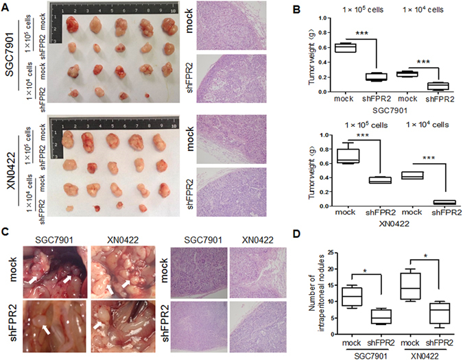
Figure 3.

FPR2 promotes tumorigenesis and metastasis of GC cells in vivo. (A) Images of xenograft tumors derived from shFPR2 SGC7901 and shXN0422 and their mock cells subcutaneously injected in nude mice at indicated cell number for 5 weeks showed that FPR2-knockdown reduced the tumorigenic abilities of GC cells both in rate of tumor formation and tumor size (left panel). HE staining confirmed the GC pathological characteristics of xenograft tumors (right panel). (B) summary graphs showed the weights of tumors derived from FPR2-knockdown SGC7901 (upper) and XN0422 (lower) cells are significantly lighter than those derived from the mock cells. (C) Representative images of intraperitoneal metastasis assay showed that metastatic nodules derived from FPR2-knockdown GC cells are far less than those derived from the mock cells (left panel), and HE staining confirms the GC pathological characteristics of the metastatic nodules (right panel). (D) Histograms show the difference in number of celiac metastatic nodules derived from FPR2-knockdown GC cells and mock cells (each cell was implanted at 2 × 10⁴ cells/mouse for 4 weeks, n = 5). *p < 0.05, ***p < 0.001.
