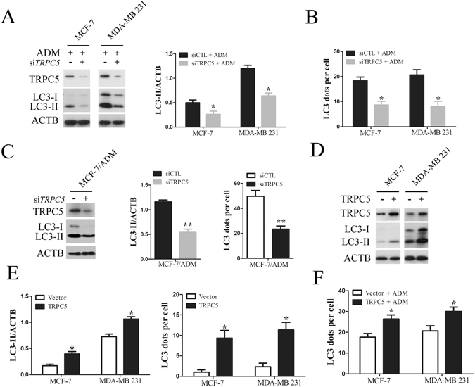
Figure 2.

Chemotherapy induced autophagy is regulated by TRPC5 in breast carcinoma cells. (A) MCF-7 and MDA-MB 231 cells were transfected with siTRPC5 or siCTL for 24 h and then exposed to ADM for 48 h. The levels of TRPC5, LC3, and ACTB were quantified by Western blot. (B) The effect of TRPC5 knockdown on the average number of LC3 dots per cell in the indicated cells. (C) Representative western blots and densitometric analysis normalized to ACTB demonstrating the effect of TRPC5 silencing on the accumulation of LC3-II and the average number of LC3 dots per cell in MCF-7/ADM cells. (D and E) Representative western blot and densitometric analysis normalized to ACTB demonstrating the effect of TRPC5 overexpression on accumulation of LC3-II and average number of LC3 dots per cell in indicated cells. (F) The effect of TRPC5 overexpression on ADM-induced LC3 puncta formation in indicated cells. Values are mean ± SEM of 3 to 6 experiments. *p < 0.05, **p < 0.01, compared to siCTL or vector.
