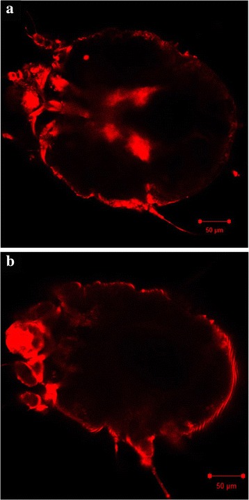
Fig. 1.

Fluorescence-labelled dsRNA uptake in Sarcoptes scabiei var. suis. Mites were immersed for 24 h at 4 °C in (a) Cy®3-labelled SsGST-mu1 dsRNA or (b) Unlabelled SsGST-mu1 dsRNA, and subsequently visualised by confocal microscopy at 20× magnification and excitation wavelength 561 nm. Scale-bars: 50 μm
