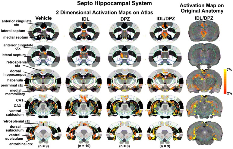
Figure 5.
Two dimensional activation maps of the septo-hippocampal system. 2D activation maps from the rat brain atlas showing the precise location of the significantly altered positive (red) voxels for each of the experimental conditions taken from the 25 to 35 min post-treatment time window in the septo-hippocampal system. The figures on the right show the localization of the voxels on the original neuroanatomical images for the combined donepezil/idalopirdine condition. The vertical color strip indicates the percent change in BOLD signal. IDL, idalopirdine; DPZ, donepezil.

