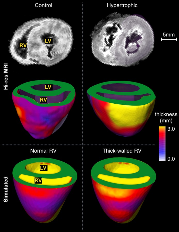
Fig. 5.

High-resolution imaging of the ventricles. Imaging from a representative control animal (left column) exhibits relatively thin RV wall compared to the LV (right column). These differences were captured by the meshes with (thick-walled RV) and without (normal) modifications in wall thickness.
