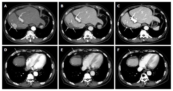
Figure 1.

Representative images of hepatocellular carcinoma spontaneously regressed. Dynamic computed tomography (CT) revealed HCC (A, arterial phase; B, portal phase) and its metastasis in the surrounding lymph node (D, arterial phase; E, portal phase). The CT images 3 mo later (C and F, arterial phase). White arrows indicate HCC and metastatic tumor in the lymph node. HCC: Hepatocellular carcinoma.
