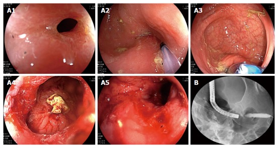
Figure 5.

Colonoscopic balloon dilatation 3 mo ago. The patient underwent X-ray-guided colonoscopic balloon dilatation at 3 mo prior to the ultimate hospital admission. Electronic colonoscopy revealed stenosis of the sigmoid colon (A1). A guide wire was inserted through the stenotic segment (A2), and a 15-mm inflatable balloon was introduced via the guide wire (A3) and gradually expanded to a diameter of 15 mm (A4 and A5). After expansion, X-ray revealed that the stenotic segment was well expanded (B).
