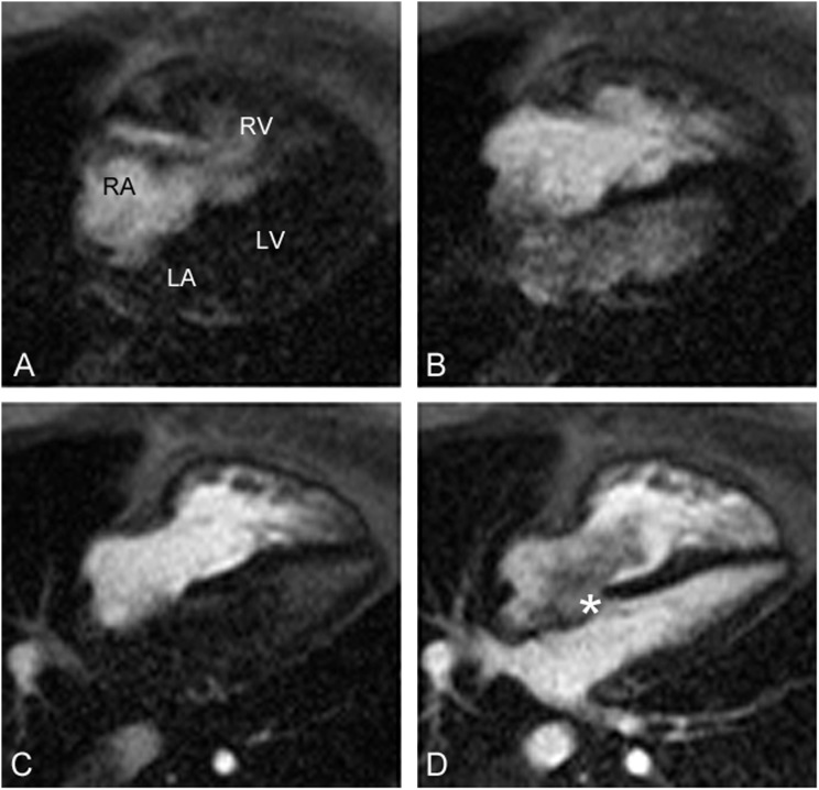
Figure 2.
Cardiac magnetic resonance imaging during injection of contrast. (A) Contrast enters the right atrium (RA) and right ventricle (RV). (B) Contrast enters the left atrium (LA) and left ventricle (LV) before entering the pulmonary veins, indicative of a right-to-left shunt at the atrial level. (C) Contrast clears from the LA and LV because of pressure equalization. (D) Contrast enters the LA and LV from the pulmonary veins. *A turbulent bidirectional shunt is present.

