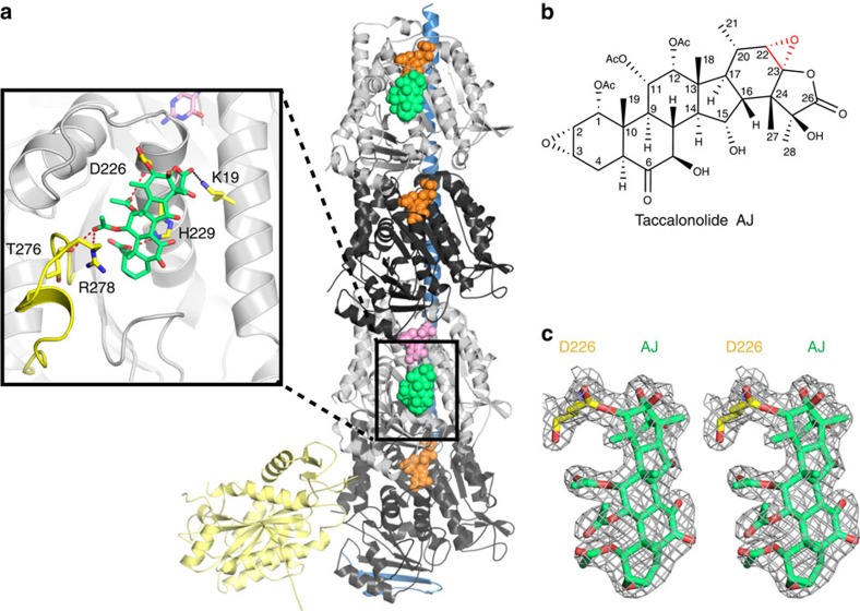
Figure 2. AJ covalent binding to β-tubulin.
(a) Overall structure of the complex crystallized. The RB3-SLD is coloured blue; TTL, light yellow; α-tubulin, black; β-tubulin, grey; GTP, orange; GDP, pink and AJ, green. The details of AJ binding are enlarged and the β M-loop is highlighted in yellow. (b) Chemical structure of taccalonolide AJ. The epoxide group between C22 and C23 is highlighted in red. (c) Stereo view of the Fo–Fc omit map (contoured at 3σ) of AJ and the side chain of βD226, clearly showing the covalent bond between them.

