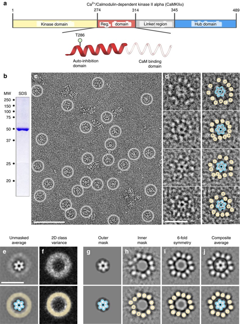
Figure 1. An extended form of the CaMKIIα holoenzyme resolved by single particle EM.
(a) Diagram of the CaMKIIα domain architecture (numbering of human isoform). Inset, shows a secondary structure diagram of the CaMKIIα regulatory domain. The coil region (faded red) represents a region that is disordered in the absence of calmodulin21. The site of auto-phosphorylation (T286) involved in regulating autonomous activity is indicated. (b) SDS–polyacrylamide gel electrophoresis of purified human CaMKIIα stained with Coomassie blue migrating at the expected molecular weight (MW ∼50 kDa). (c) Electron micrograph of negatively stained CaMKIIα particles. Contrast of protein is white on a dark background. Individual particles are indicated by white circles. Scale bar=100 nm. (d) Enlarged view of individual particles with the hub complex (blue outline) and twelve kinase domains (yellow circle) indicated. Scale bar=25 nm. (e,f) Single particle averaging and image analysis. Scale bar=25 nm. (e) Representative 2D projection average of unmasked particles. The central hub complex is clearly defined (blue outline), while the radial kinase domains appear as a diffuse halo (yellow circle). (f) 2D variance map of data in e. A region of high variance (white pixels) corresponds with the blurred region observed in 2D class averages (yellow circle). (g) Representative 2D projection average using an applied image mask (75 Å outer radius). The sixfold symmetric hub complex is clearly defined (blue outline). (h) Representative 2D projection average with an applied mask to remove contribution of the hub domain during the alignment procedure (50 Å inner radius mask). Twelve kinase domains are visualized at an approximate radius of 24–28 nm (yellow circles). (i) Class average in h with applied sixfold rotational averaging indicates a pseudo-symmetric kinase domain organization. (j) Composite image of g and i with the identified hub complex (blue outline) and kinase domains (yellow outline) indicated.

