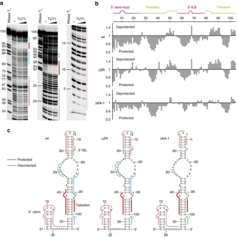
Figure 4. Interaction of human TUT1 with U6 snRNA.
(a) Tb(III)-mediated cleavage pattern of U6 snRNA in the absence and presence of TUT1. 32P-labelled U6 snRNA-u4 was incubated with Tb(III) in absence or presence of 0.4 μM and 0.8 μM recombinant TUT1. Positions of nucleotides were determined by partial digestion of the RNA substrate by RNase T1. Cleavage patterns were analysed on 8% (w/v) (left) or 16% (w/v) (middle and right) sequencing gels. Protected and deprotected regions are depicted by red and cyan lines, respectively. (b) Quantitative analysis of TUT1 binding to U6 snRNA. The relative band intensities at each nucleotide position, in the presence and absence of wild-type TUT1 (upper), are shown. Quantitative analysis of ΔZR (middle), and ΔKA-1 (lower) binding, as in wild-type TUT1 (Supplementary Figs 8,9). (c) Superimpositions of the footprinting data of wild-type TUT1 (left), ΔZR (middle) and ΔKA-1 (right) onto the secondary structure of the U6 snRNA34. Protected and deprotected regions are coloured as in a.

