Fig. 3.

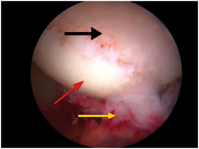
Arthroscopic image of articular changes in the femoral head (black arrow) with an adjacent normal articular surface (red arrow) and a torn and degenerate LT (yellow arrow).

Arthroscopic image of articular changes in the femoral head (black arrow) with an adjacent normal articular surface (red arrow) and a torn and degenerate LT (yellow arrow).