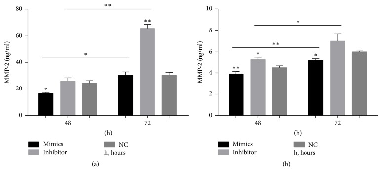
Figure 5.
Changes in matrix metalloproteinase- (MMP-) 2 secretion from scleral fibroblasts and retinal pigment epithelial (RPE) cells after transfection with miR-29a mimics or inhibitor. The protein secreted from scleral fibroblasts and RPE cells was analyzed using enzyme-linked immunosorbent assays (ELISAs). The secretion of MMP-2 by scleral fibroblasts after 48 hours was decreased in cells transfected with the miR-29a mimics compared with negative controls (P < 0.05). The secretion of MMP-2 by scleral fibroblasts after 72 hours was significantly increased in cells transfected with the miR-29a inhibitor compared with negative controls (P < 0.01). The secretion of MMP-2 was higher 72 hours after transfection than 48 hours after transfection in both the miR-29a mimics group (P < 0.05) and miR-29a inhibitor group (P < 0.01). (a) The secretion of MMP-2 by RPE cells was significantly decreased after 48 (P < 0.01) and 72 (P < 0.05) hours in cells transfected with the miR-29a mimics compared with negative controls. The secretion of MMP-2 by RPE cells after 48 hours was significantly increased in cells transfected with the miR-29a inhibitor compared with negative controls (P < 0.05). The secretion of MMP-2 was higher 72 hours after transfection than 48 hours after transfection in both the miR-29a mimics group (P < 0.01) and miR-29a inhibitor group (P < 0.05) (b). Note: the error bars show the standard deviations (n = 3). The statistical analyses were performed using one-way ANOVA for groups at same time point and paired t-tests for between-group analyses at different time points. ∗P < 0.05, ∗∗P < 0.01. NC, negative control; h, hour.

