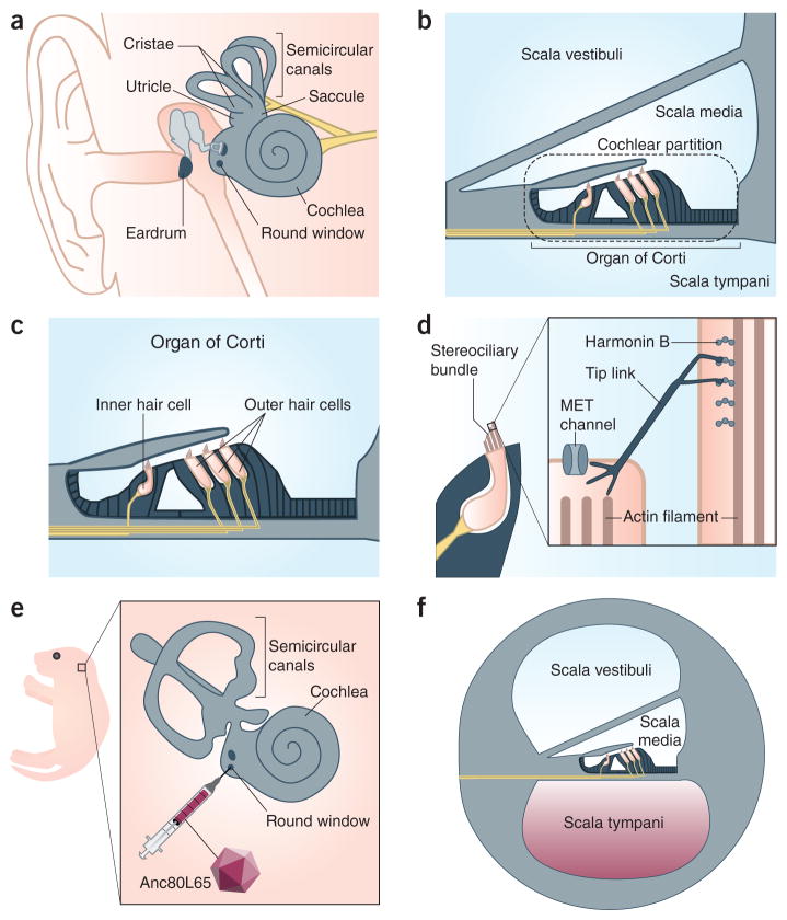
Figure 1.
Structure, function, and viral transduction of the inner ear. (a) The human inner ear. The vestibular sensory epithelium is located in the cristae of the three semicircular canals and the maculae of the utricle and saccule. The auditory sensory epithelium is contained within the coiled cochlea. The round window is covered by an uncalcified membrane. (b) The three fluid-filled compartments of the inner ear are called the scalae. Inner and outer sensory hair cells are in the organ of Corti. The cochlear partition oscillates in response to vibrations produced in the scala media by movement of the ear drum. (c) The organ of Corti within the cochlea presents one row of inner hair cells and three rows of outer hair cells which are surrounded by supporting cells (black). (d) The stereociliary bundle on the apical surface of hair cells consists of parallel stereocilia whose structural rigidity is established in part by filamentous actin. A proteinaceous tip link connects adjacent stereocilia and has an important role in the activation of the mechanoelectrical transduction (MET) channel. Harmonin-b is localized to the upper insertion point of the tip link, where it interacts with numerous proteins. (e) A postnatal day 1 mouse pup whose left inner ear (inset) is targeted for round window membrane injection with Anc80L65. (f) The solution containing Anc80L65 fills the scala tympani and then spreads to the other two scalae, gaining access to structurally and functionally immature neonatal hair cells.

