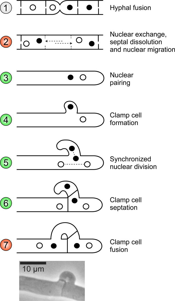
Fig. 5. Roles of P/R and HD genes on the formation and maintenance of the dikaryon in C. cinerea.

Pheromone signaling is not required to attract mates and hyphal fusion is mating-type independent (diagram 1). Upon fusion, nuclei enter the mycelium of the other mate and migrate until they reach a hyphal tip cell (diagram 2). During hyphal tip elongation, the two types of haploid nuclei (depicted in white and black, representing different MAT genotypes) pair at the tip cell (diagram 3) and, at the place where mitosis will take place, a hook-like structure (called clamp connection) is formed (diagram 4). The two nuclei divide synchronously: one of the nuclei divides in direction of the clamp cell that is growing backwards toward the main hyphae, while the other divides along the main hyphal axis (diagram 5). Septa are generated between the dividing nuclei. This way one nucleus stays temporarily entrapped into the clamp cell, one nucleus of the other type is enclosed in the newly formed subapical cell, and a nucleus of each type is maintained in the emerging hyphal tip cell (diagram 6). The clamp cell fuses with the subapical cell and releases the entrapped nucleus from the clamp cell, restoring the dikaryotic state of the subapical cell (diagram 7). In C. cinerea, steps controlled by P/R (diagrams 2 and 7) and HD (diagrams 3 to 6) genes are colored in red and green, respectively. See Casselton et al. (18) and Kües (90) for details. Micrograph at the bottom were obtained from Stajich et al. (86), with permission.
