
Figure 4.
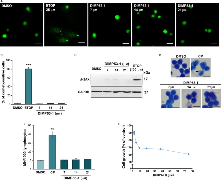
DIMP53‐1 is nongenotoxic in normal and tumor cells and has low cytotoxicity against normal cells. (A–C) DNA damage was measured in HCT116p53+/+ cells by comet assay (A and B) and by analysis of γH2AX expression levels (C) after treatment with etoposide (ETOP; positive control) or DIMP53‐1. In (A), scale bar = 20 μm; magnification = 200 ×. In (B), quantification of comet‐positive cells (containing more than 5% of DNA in the tail; assessed by open comet/imagej); 100 cells were analyzed in each group. In (C), γH2AX levels were determined by western blot; immunoblots are representative of three independent experiments; GAPDH was used as loading control. (D and E) Genotoxicity of 7, 14, and 21 μm DIMP53‐1 by cytokinesis‐block micronucleus (MN) assay after 72‐h treatment in human lymphocyte cells; 5 μg·mL−1 cyclophosphamide (CP; positive control). In (D), scale bar = 20 μm; magnification = 1000 ×. In (E), the number of MN per 1000 binucleated lymphocytes was recorded. (F) DIMP53‐1 concentration–response growth curve in MCF10A cells, after 48‐h treatment; incubation with DMSO, in equivalent % of DIMP53‐1, was used to normalize the results. In (B), (E), and (F), data are mean ± SEM of three to four independent experiments; in (B) and (E), values were significantly different from DMSO (**P < 0.01; ***P < 0.001).