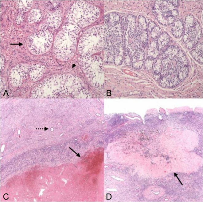
Figure 2.

Ovo-testis histology. (A) Seminiferous tubules (arrow), devoid of germ cells or spermatogenesis, with interstitial Leydig cells (arrow head). (B) Sertoli cell nodule in testicular component (Leydig-rich background). (C) Ovarian tissue with an involuting haemorrhagic corpus luteum (arrow) and a primordial follicle (dotted arrow). (D) Ovarian tissue with an old corpus albicans (arrow), the adjacent ovarian stroma with a further primordial follicle. Image dimensions, A and B: 0.9 × 0.7 mm; C and D: 2.2 × 1.7 mm.

 This work is licensed under a