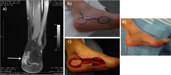
Fig. 5.
This 16- year-old male had a lump resection of the heel, without pre-operative imaging or biopsy. The diagnosis was a clear cell sarcoma. a) MRI of the left foot before ‘second-look’ surgery (T1 fat sat sequence coronal view). b) Drawing of the planned resection and flap. c) Intra-operative view after resection of the heel tumour and rotation of the medial plantar flap. Clear margins were achieved. d) Appearance of the foot two years after surgery.

