
Figure 1.
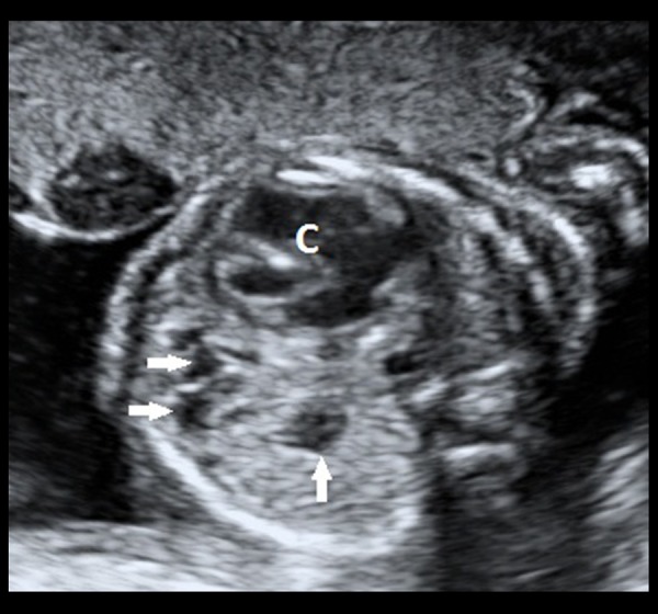
Grey-scale transabdominal ultrasound image of the foetus in a transverse section showing multiple, anechoic structures (white, filled arrows) within the lower lobe of the left lung. The cardia (denoted by ‘C’) appears to be mildly pushed to the right side.