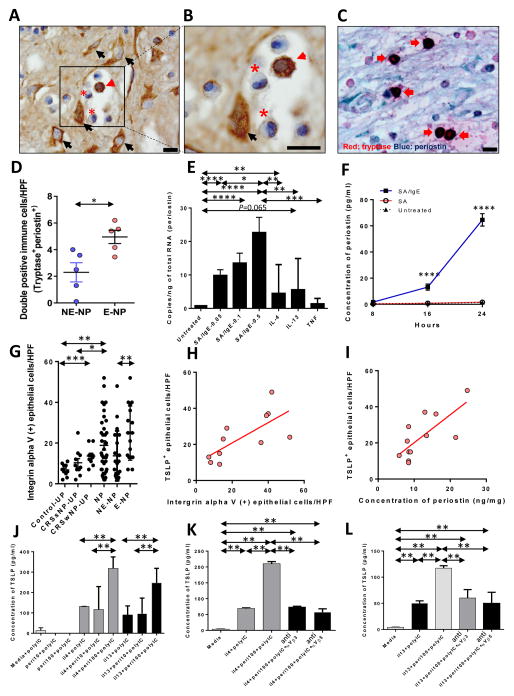
Figure 1. Cross-talk between human mast cells and epithelial cells in eosinophilic nasal polyps.
(A, B) IHC detection of periostin was performed within the NP tissues from patients with CRSwNP (eosinophil, asterisk; mast cell, red arrow head; fibroblast, black arrow). Black scale bar=10μm. (C) Representative double immunostaining (red: tryptase and blue: periostin). Arrows indicate double-positive immune cells (x1000). Black scale bar=10μm. (D) Counts of double-positive cells in NE-NP and E-NP (n=5 for each group). (E) Periostin mRNA expression was measured by qRT-PCR in LAD2 cells stimulated with IgE, IL-4, IL-13 and TNF-α (n=9 for each group). (F) Periostin protein expression stimulated by IgE (n=3 for each group). (G) Number of integrin αV-positive epithelial cells per 100 cells was determined and averaged from three different areas of epithelium (n=10 for Control-UP, n=10 for CRSsNP-UP, n=10 for CRSwNP-UP, and n=37 for CRSwNP-NP) in IHC assays of tissue sections. The number of integrin αV-positive inflammatory cells was counted from five of the densest areas (HPF; x400) and averaged for each group. (H) Correlation between integrin αV and TSLP at the protein level. (I) Correlation between periostin and TSLP at the protein level. (J) Periostin-induced TSLP production in NHBE cells after Th2 cytokine and Poly(I:C) treatment (n=3 for each group). (K, L) Periostin-induced TSLP production was reversed by anti-integrin αVβ3 (K) and anti- integrin αVβ5 (L) (n=3 for each group). *P<0.05, **P<0.01, ***P<0.001, and ****P<0.0001 using the Mann-Whitney U-test.

