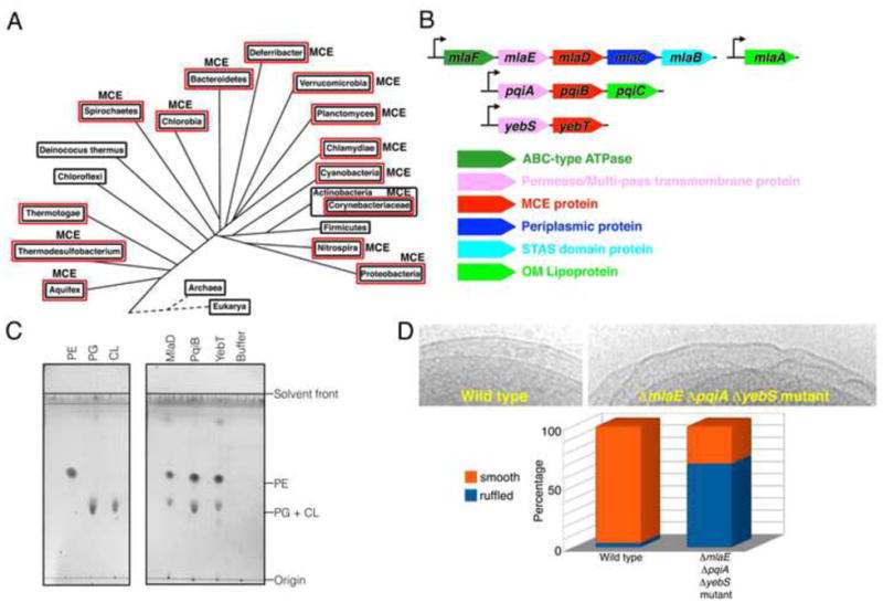
Figure 1. MCE proteins are important for OM integrity.

(A) Distribution of MCE proteins across bacterial lineages. Red boxes indicate double-membranes.
(B) Organization of MCE operons from E. coli.
(C) TLC of phospholipids bound to purified E. coli MCE proteins. Left, lipid standards. Right, lipids co-purifying with MlaD, PqiB, and YebT.
(D) OM ruffling of MCE mutants. Top, electron micrographs of WT and ΔmlaE ΔpqiA ΔyebS E. coli cells, with smooth and ruffled OM, respectively. Bottom, frequency of OM ruffling (n~50 cells).
