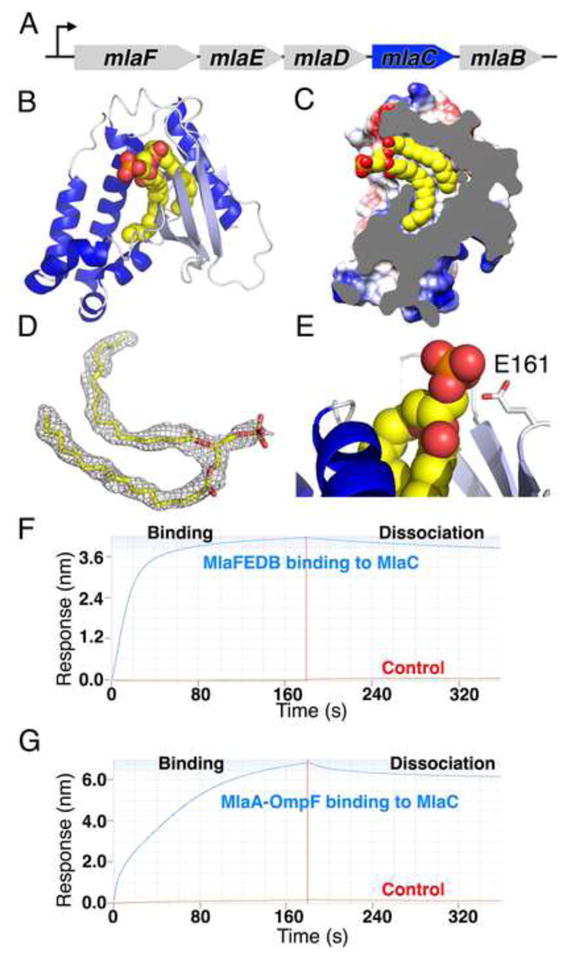
Figure 4. MlaC shuttles lipids across the periplasm.

(A) MlaC is encoded in the mla operon but not stably present in the MlaFEDB complex.
(B) Crystal structure of MlaC reveals a mixed α/β fold with 4 β-strands (light blue) and 7 α-helices (dark blue). MlaC-bound lipid shown as spheres.
(C) Cut away view of MlaC surface, revealing deep hydrophobic pocket that binds the two “tails” of a lipid.
(D) Unbiased 2Fo-Fc electron density map for bound lipid, contoured at 1 σ.
(E) MlaC makes no significant interactions with the lipid head group.
(F and G) MlaC interacts with the IM MlaFEDB complex (F) and the OM MlaA-OmpF complex (G), suggesting MlaC may ferry lipid substrates between these IM and OM complexes.
