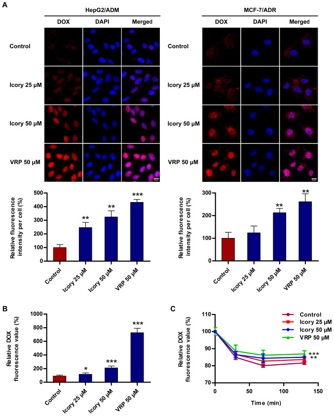
Figure 4.
Icory increases the accumulation of DOX and inhibits the efflux of DOX in HepG2/ADM cells. (A) Cells were observed by fluorescence microscopy, and the representative images (top) and quantitative analysis (bottom) of DOX fluorescence are shown. DOX staining are shown in red. DAPI (blue) counter-stains the nuclei. Original amplification: 630; bar: 10 μm. (B) The intracellular accumulation of DOX was measured with a DTX 880 Multimode Detector. (C) The effect of Icory on DOX efflux in HepG2/ADM cells. The intracellular DOX level was measured using a DTX 880 Multimode Detector in different periods of time (0, 30, 70 and 130 min). VRP (50 μM) was used as a positive-control inhibitor of ABCB1. The results are presented as the means ± SD of three independent experiments. *P<0.05, **P<0.01 and ***P<0.001 compared with the control.

