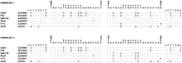
FIG. 1.
Comparison of partial phlD sequences from several Pseudomonas strains. Bases that are identical in all sequences are indicated by dashes. The sequences and positions of the two primer sets used in PCR-DGGE analysis are shown above each of the two alignments. The positions of the 5′ and 3′ ends of each of the primers correspond to the positions in the phlD sequence of Pf-5 (accession number AF214457).

