
Figure 3.
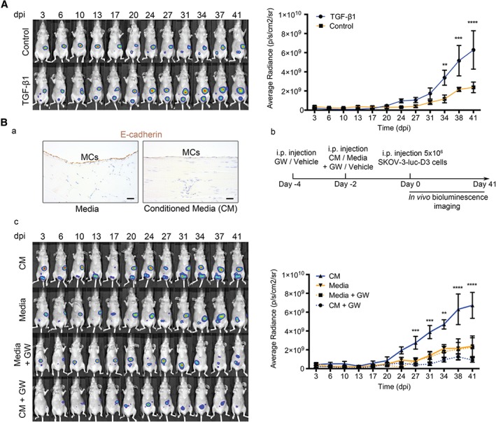
OvCa‐secreted TGF‐β transforms the pre‐metastatic peritoneum, favouring tumour progression. (A) Representative images of in vivo monitoring of SKOV3‐luc‐D3 cells in mice pre‐conditioned with TGF‐β1‐encoding adenovirus or control. Quantification of bioluminescence showed that tumour growth was increased in mice pre‐conditioned with TGF‐β1 adenovirus (n = 6) as compared with control adenoviral pretreatment (n = 6). (B) (a) Representative images of E‐cadherin immunostaining in the mesothelial monolayer of a mouse killed 2 days after being pre‐conditioned with conditioned medium (CM) from OvCa cells or control medium. Scale bars: 25 µm. (b) Diagram of experimental design. (c) Representative images of in vivo monitoring of SKOV3‐luc‐D3 cells and quantification of bioluminescence showed that intraperitoneal tumour growth was higher in mice pretreated with SKOV3 medium (CM). The TGF‐β receptor I inhibitor (GW) reduced tumour growth to levels comparable to those of mice whose peritoneums had not been pre‐conditioned (control medium). n = 6 per group. All mice were monitored for 41 days. Graphs represent mean average radiance (expressed as photons/s/cm2/sr) of SKOV3‐luc‐D3 cells ± standard error of the mean. Symbols represent the statistical differences over time between groups (**p ≤ 0.01; ***p ≤ 0.001; ****p ≤ 0.0001). dpi, days post‐inoculation; i.p., intraperitoneal.