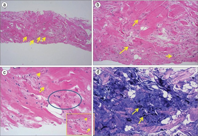
Figure 3.
(a, b) Hematoxylin and eosin staining of the biopsy specimen shows altered elastin fibers (yellow arrows) and interspersed mature adipose tissue in a fibrous background. (c) Note the characteristic wavy, serrated edges (yellow arrow) and beaded appearance (blue circle) of the elastic fibers. The inset shows a magnified view. (d) Elastin stains show deeply staining branched and unbranched elastin fibers (yellow arrows).

