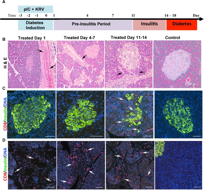
Fig 1. Pancreas inflammation in diabetes-induced rats occurs prior to insulitis while islet morphology is normal.
(A) Pancreas tissue was collected from pIC+KRV treated and control rats at the time points indicated and processed for histology and immunostaining. (B) Representative images of H&E stained pancreas sections from pIC+KRV treated and control rats. n = 6 rats/time point; scale bars, 100 um. Arrows indicate vessel/duct dilation. Representative immunofluorescent images of islets (C) and exocrine tissue (D) from pancreas sections of pIC+KRV treated and control rats; CD8 (red), insulin (green), DNA (blue). n = 4 rats/time point; scale bars, 50 um. Arrows indicate CD8+ cells.

