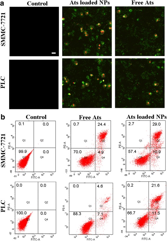
Fig. 5.

Morphology of the ultrastructural changes of cells treated with free Ats and Ats-loaded BSA NPs using Annexin V-FITC/PI staining assay (a). Scale bar, 100 μm. Flow cytometer analysis of the cellular apoptosis and oncosis after 24 h of incubation with the free Ats and Ats-loaded BSA NPs, respectively (b)
