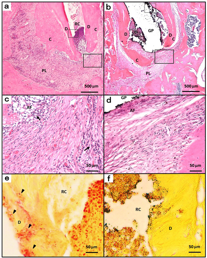
Figure 6.

Histologic images at week 12. (a) Periapical areas of the control group stained with haematoxylin and eosin. (b) Periapical areas of the treatment group stained with haematoxylin and eosin. (c and d) High-magnification views of the solid inset in panels a and b, respectively. (e and f) High-magnification views of the dotted insets in panels b and e, respectively, stained with a modified Brown and Brenn method. AF, apical foramen; GP, gutta-percha point; RC, root canal; C, cementum; D, dentin; PL, periapical lesion; arrow head, bacteria; arrow, inflammatory cells.
