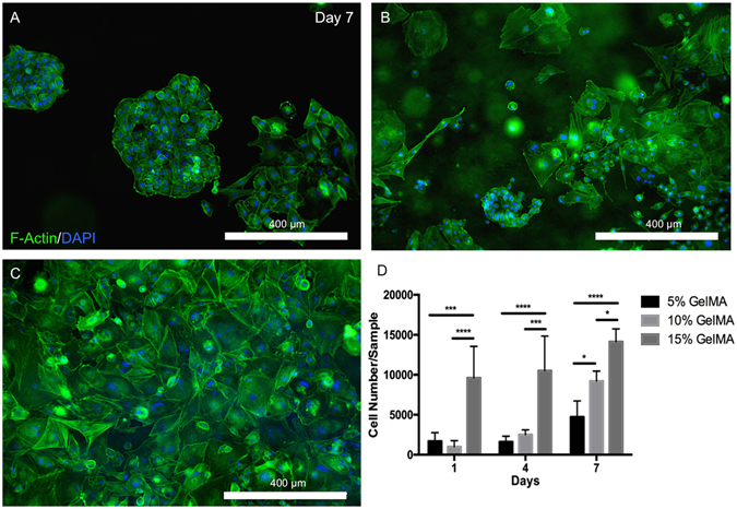
Figure 4.

Spreading and proliferation of OD21 cells in 3D GelMA hydrogels. Representative images of OD21 cells encapsulated in (A) 5%, (B) 10% and (C) 15% GelMA hydrogels stained for actin (green) and DAPI (blue) shows increased cell spreading in stiffer over softer hydrogels. (D) Quantification of the number of cells per gel after 1, 4 and 7 days in culture indicates a significantly higher rate of proliferation of OD21 cells in scaffolds of higher polymer concentration. Statistical significance is represented by *for p < 0.05, ***for p < 0.001 and ****for p < 0.0001.
