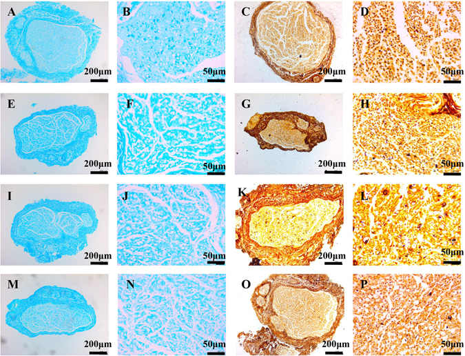
Figure 1.

Histopathological examination of injured facial nerves on day 7 by Luxol fast bule staining (A,B,E,F,I,J,M,N) and Bielschowsky staining (C,D,G,H,K,L,O,P). Grade I injured facial nerve (A-D) had an integrated perineurium and continuous axons with well-distributed and aligned myelin sheaths in the cross-sections. Grade II injured facial nerve (E–H) had patchy loss of myelin sheaths with intact nerve bundles. Grade III injured facial nerve (I–L) had substantial thinning of myelin sheaths and blistered and vacuolar degenerative axons. Most of the myelin sheaths and axons were disintegrated in Grade IV injured facial nerve (M–P).
