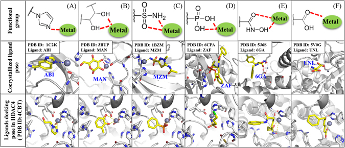
Figure 3.

Validation of metal binding groups in HDAC. Structure of metal binding group for imidazole (A), diols (B), sulfonamids (C), phosphonates (D), hydroxamic acids (E), and carboxylic acids (F) located at top. The middle panel show structures of co-crystallized ligands with functional groups. The PDB ID of ligands are shown in blue. The small fragment docking docked in HDAC4 crystal structure is shown at the bottom. Grey sphere represents zinc ion.
