
Figure 3.
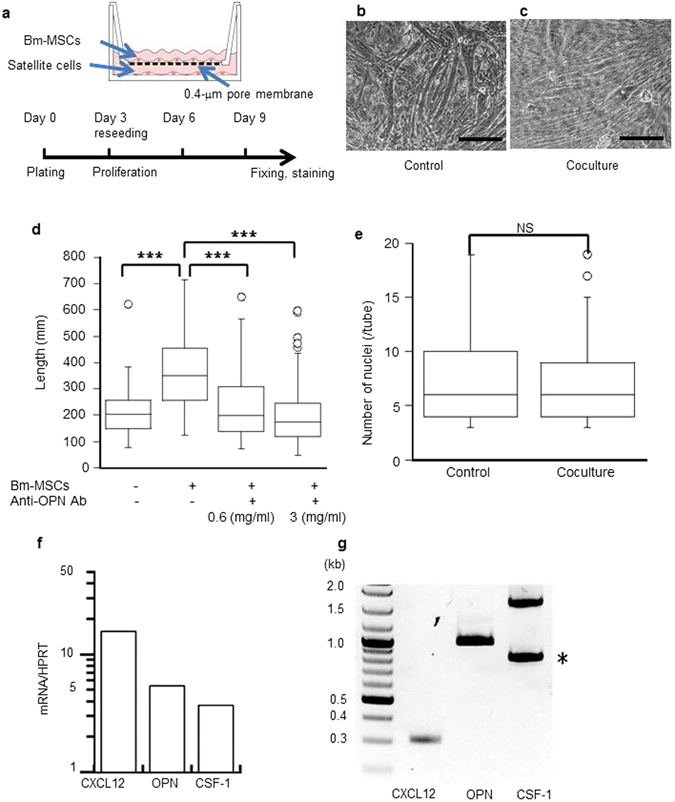
Bm-MSCs produced various growth factors and affected in vitro myotube formation. (a) Bm-MSCs (1.0 × 10⁵ cells per insert) were added to culture inserts 1 day before starting cocultures with single myofibers isolated from the calf muscle of the wild-type mouse. The culture design is described in detail in Material Methods. Micrographs showing that, compared with the control culture (b), myotubes cocultured with Bm-MSCs (c) were longer and grew in a definite direction. Scale bars: 100 μm. (d) Addition of anti-OPN antibody to the coculture inhibited these effects in an antibody concentration-dependent manner. Myotube length: control, 216 ± 93.3 μm; coculture, 369.7 ± 174.5 μm (***P < 0.001; n = 65–75 in each group). Data represent at least three independent experiments. (e) The number of nuclei in each myotube in the control and coculture groups did not differ. This result suggests that the fusion frequency of the myoblasts was the same. NS, not significant. (f) The mRNA-PCR result showing that Bm-MSCs expressed high amounts of CXCL12, OPN, and CSF-1. HPRT, hypoxanthine-guanine phosphoribosyltransferase. (g) RT-PCR showing full-length CXCL12, OPN, and CSF-1 in Bm-MSCs. *Indicates a fragmentation of OPN.